Mesothelioma Histology Microvilli / Pathology Outlines Mesothelioma Peritoneum Epithelioid / It labels normal mesothelial cells as well as epithelial mesotheliomas in a thick membranous pattern ( dahlstrom et al., 2001 ;
Mesothelioma Histology Microvilli / Pathology Outlines Mesothelioma Peritoneum Epithelioid / It labels normal mesothelial cells as well as epithelial mesotheliomas in a thick membranous pattern ( dahlstrom et al., 2001 ;. The aetiology of this neoplasm in the tunica vaginalis is not known. Quantitative evaluation of the length to diameter ratio of the 10 longest microvilli revealed a mean value of approximately 16.2. Numerous microvilli seen by electron microscopy. mesothelioma is an important cause of lobulated or nodular pleural thickening (fig. Extremely early age of onset (7 weeks old) suggests that congenital mesothelioma may occur infrequently in the dog.
Leathers mesotheliomas are rare neoplasms. It stains normal mesothelial cells as well as epithelial mesotheliomas in a thick membrane pattern. Differentiation from adenocarcinoma was based on ultrastructural features, particularly on the morphology of the microvilli and the presence of microfilaments. However, documented cases of mesothelioma have been reported in patients from as young as 7 weeks to as old as 15 years. Primary tumors arising from mesothelium of the peritoneum are extremely rare.
Malignant Mesothelioma And Other Mesothelial Proliferations Chapter 28 Modern Soft Tissue Pathology from static.cambridge.org Quantitative evaluation of the length to diameter ratio of the 10 longest microvilli revealed a mean value of approximately 16.2. Chapter 79 malignant mesothelioma and other primary pleural tumors edmund k. There were four women and two men, ranging in age from 42 to 76 years. Morbidity, mortality, mean survival, and the impact of histology on survival after pleurectomy in 64 patients with malignant pleural mesothelioma. The detection of long microvilli, an ultrastructural feature unique for malignant epithelial mesothelioma, requires immunolabeling of epithelial membrane antigen to be visible by light microscopy. mesothelioma cells had longer, thinner microvilli on the cell surfaces (p < The epithelioid mesothelioma cells displayed long microvilli, cytoplasmic. microvilli but not long and slender.
microvilli are characteristic of mesothelial cells.
The epithelioid mesothelioma cells displayed long microvilli, cytoplasmic. Summarized, this report described the case of a malignant biphasic mesothelioma with an atypical ck20 expression but a characteristic ultrastructural morphology including long microvilli. There were four women and two men, ranging in age from 42 to 76 years. mesothelioma (mpm) is a tumour rind that follows the contour of the inner chest wall to encircle the lung, in microvilli characteristic of mpm, and in 9 of 20 cases of lung adenocarcinomas (45%), cells with short microvilli were evidenced. Tem ultrastructure of cultured mesothelioma cells showed microvilli of various lengths in different cell lines, but overall the microvilli in cultured mesothelioma cells were shorter than those observed in histological samples of mesothelioma. Ultrastructurally, the tumor cells showed long slender microvilli on the apical surface, consistent with mesothelioma. The article deals with cytopathology specimens from spaces lined with mesothelium, i.e. Immunohistochemically, the various mesothelial markers were positive, and a few adenocarcinoma markers were focally positive. However, this test can be positive in almost any malignancy and, thus, an immunohistochemical panel to prove mesothelial origin should still be the first step in diagnosis. Occasional cells have intracytoplasmic vacuoles. mesothelioma with a pronounced myxoid stroma has been identified as a morphological pattern that might portend a better prognosis. Of histology, histochemistry, immunohistochemistry, and, if possible, electron microscopy.
The epithelioid mesothelioma cells displayed long microvilli, cytoplasmic. The team showed that microvilli — tiny moving protrusions that help the lungs to expel dust, mucus, and microbes — had particular features in the two cancers. Of histology, histochemistry, immunohistochemistry, and, if possible, electron microscopy. Learn vocabulary, terms, and more with flashcards, games, and other study tools. The article deals with cytopathology specimens from spaces lined with mesothelium, i.e.
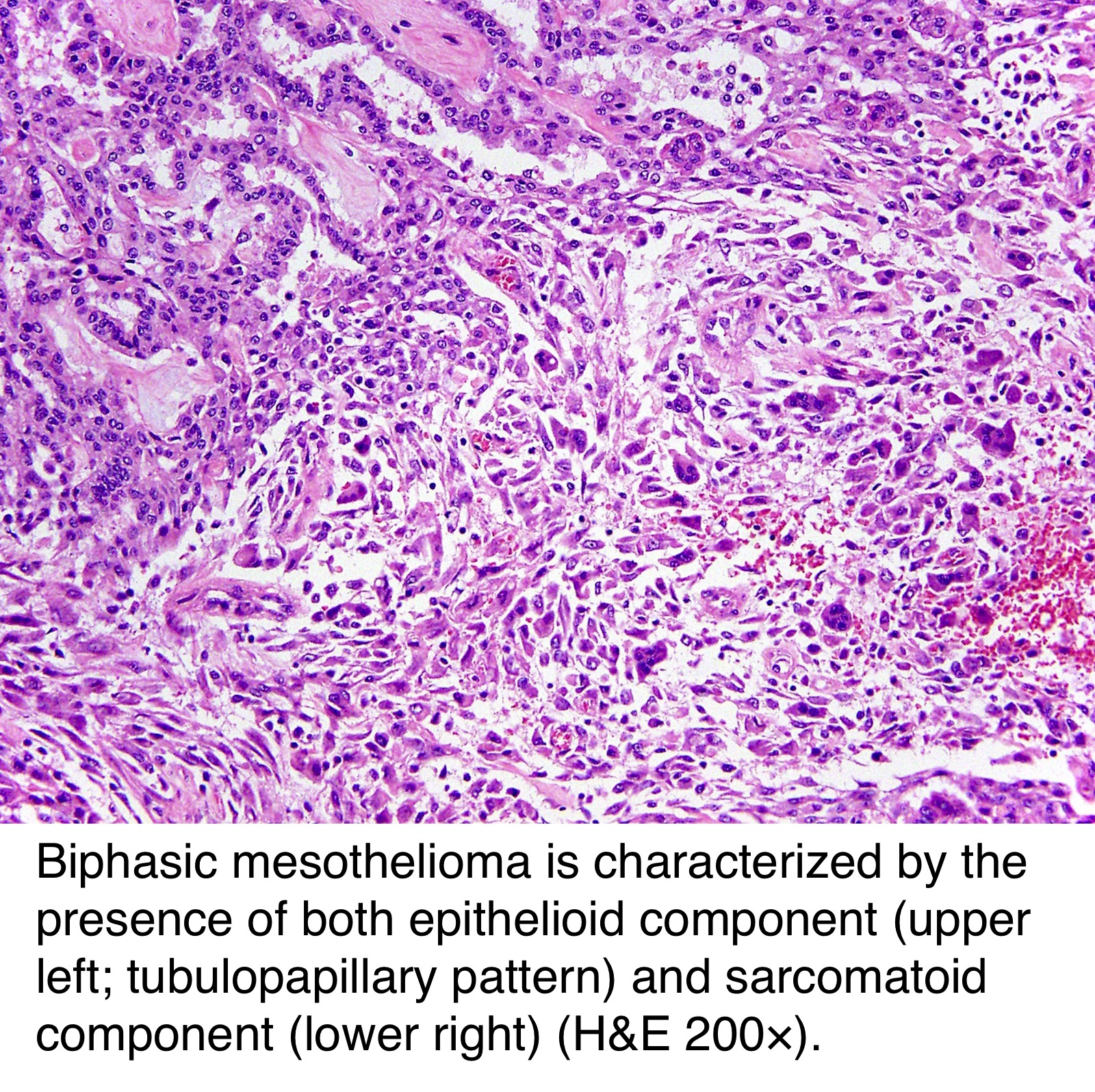
Pathology Outlines Peritoneal Malignant Mesothelioma from www.pathologyoutlines.com Remember that some primary pulmonary adenocarcinomas have long microvilli, but these microvilli are invariably covered by a glycocalyx. microvilli are characteristic of mesothelial cells. mesothelioma cells had longer, thinner microvilli on the cell surfaces (p < mesothelioma cells had long, thin microvilli that lacked the outermost layer — called the glycocalyx — needed for the attachment of substances to the protrusions. A proper diagnosis of biphasic mesothelioma involves first distinguishing between mesothelioma and other types of cancer that have biphasic characteristics such as synovial sarcomas and. This is an unusual lesion in which the neoplastic cells are separated from each other by abundant stromal mucin. The article deals with cytopathology specimens from spaces lined with mesothelium, i.e. Short and plump microvilli found in adenoca/mesothelioma.
Histopathological studies can only be conducted with a certified and experienced medical professional.
The detection of long microvilli, an ultrastructural feature unique for malignant epithelial mesothelioma, requires immunolabeling of epithelial membrane antigen to be visible by light microscopy. Immunohistochemically, the various mesothelial markers were positive, and a few adenocarcinoma markers were focally positive. 3.10).124,131,239124131239 mesothelioma spreads around the pleura and is virtually always unilateral. Deciduoid mesothelioma is a rare variant of epithelial mesothelioma, up to now only described in human pathology, which bears remarkable cytomorphologic resemblance to the endometrium of pregnancy, termed decidua. At histology, it was observed a neoplastic proliferation morphologically compatible with mesothelioma with several foci of squamous differentiation (figure 1a,b). mesothelioma of pleura with long, curved microvilli. mesothelioma cytology or mesothelioma cytopathology is the study of cells for the presence of mesothelioma. Short and plump microvilli found in adenoca/mesothelioma. mesothelioma with a pronounced myxoid stroma has been identified as a morphological pattern that might portend a better prognosis. Chapter 79 malignant mesothelioma and other primary pleural tumors edmund k. Fine needle aspiration (fna) of this type of tumor has rarely been reported. The aetiology of this neoplasm in the tunica vaginalis is not known. Immunohistochemical reactions were positive for markers of mesothelial origin ( figure 1 c,d), while in the squamous component, the neoplastic cells strongly expressed p40 ( figure 1 e).
However, documented cases of mesothelioma have been reported in patients from as young as 7 weeks to as old as 15 years. Carcinoma, including secretory granules, and lacked typical features of mesothelioma, such as long slender microvilli. No distinctive histologic pattern some are sinusoidal or have fibrillar matrix; Sarcomatoid mesothelioma (sm) is the rarest subtype of malignant mesothelioma (mm). The epithelioid mesothelioma cells displayed long microvilli, cytoplasmic.
What Is Mesothelioma Histology Variances In Histological Mesothelioma from dekorasikartini.com It labels normal mesothelial cells as well as epithelial mesotheliomas in a thick membranous pattern ( dahlstrom et al., 2001 ; Of histology, histochemistry, immunohistochemistry, and, if possible, electron microscopy. Histopathological studies can only be conducted with a certified and experienced medical professional. Eight cases of mesothelioma with small cell features were identified from a review of 960 cases of mesothelioma from the files of the department of pathology at the university of texas md anderson. Sterman the pleura is a membranous structure covering the entire surface of the lung and lining the inside of the chest cavity. A general differential diagnosis of pleural effusion is given. In contrast to previously reported cases of deciduoid mesothelioma, this tumor developed in the abdominal wall and appears to have a benign course. mesothelioma cells had long, thin microvilli that lacked the outermost layer — called the glycocalyx — needed for the attachment of substances to the protrusions.
However, this test can be positive in almost any malignancy and, thus, an immunohistochemical panel to prove mesothelial origin should still be the first step in diagnosis.
mesothelioma of pleura with long, curved microvilli. The aetiology of this neoplasm in the tunica vaginalis is not known. The detection of long microvilli, an ultrastructural feature unique for malignant epithelial mesothelioma, requires immunolabeling of epithelial membrane antigen to be visible by light microscopy. The epithelioid mesothelioma cells displayed long microvilli, cytoplasmic. microvilli but not long and slender. Occasional cells have intracytoplasmic vacuoles. Eight cases of mesothelioma with small cell features were identified from a review of 960 cases of mesothelioma from the files of the department of pathology at the university of texas md anderson. While it has not so far proven to be a useful diagnostic determinant to indicate whether a patient is suffering from mesothelioma, it has been extremely useful in the differential diagnosis of mesothelioma versus adenocarcinoma or other similar types of cancer. It labels normal mesothelial cells as well as epithelial mesotheliomas in a thick membranous pattern ( dahlstrom et al., 2001 ; Of the 64 cases of deciduoid mesothelioma, including the 43 reported in the literature ( table 1) and the 21 in the present series ( table 3 ), 34 occurred in males who ranged in age from 13 to 78. The study of diseased cells, such as those found in tumors, is a branch of histology called histopathology. The diagnosis can pose several problems to the At histology, it was observed a neoplastic proliferation morphologically compatible with mesothelioma with several foci of squamous differentiation (figure 1a,b).
The study of diseased cells, such as those found in tumors, is a branch of histology called histopathology mesothelioma histology. At histology, it was observed a neoplastic proliferation morphologically compatible with mesothelioma with several foci of squamous differentiation (figure 1a,b).


0 Comments